|
| | July 31, 2019 | |
| | |
| | The latest life science microscopy news from AZoNetwork | |
|
|
 |
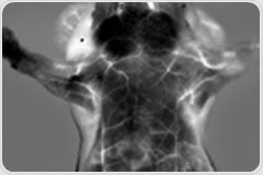
| |  NIR-II Fluorescence Imaging - Opening a New Window into Living Tissues for Preclinical Research NIR-II Fluorescence Imaging - Opening a New Window into Living Tissues for Preclinical Research
Taking advantage of increased penetration and contrast when imaging tissues in the NIR-II, Photon etc.’s webinar will be bridging the gap between radiology and pathology. Reaching a penetration depth of up to 3 cm in a mouse enables the high-resolution visualization of full organs and microvasculature while monitoring blood flow, metabolic rates, cellular environment, perfusion, drug biodistribution, tumour progression, heart and respiratory rates.
| |
|
|
|
|
 |
| | | |
|
| |  Nanotechnology-based compounds are now being tested as a possible delivery vehicle for vaccines. A team of researchers demonstrated how the oral delivery vehicle SBA-15, one such nanotechnology-based compound can deliver an oral vaccine against hepatitis B to the immune system. Nanotechnology-based compounds are now being tested as a possible delivery vehicle for vaccines. A team of researchers demonstrated how the oral delivery vehicle SBA-15, one such nanotechnology-based compound can deliver an oral vaccine against hepatitis B to the immune system. | |
|
| |  E-FRET microscopy recently became a very popular intensity-based Fluorescence Resonance Energy Transfer (FRET) method for cell quantification. This technique reduces the photobleaching observed during the process of FRET. E-FRET microscopy recently became a very popular intensity-based Fluorescence Resonance Energy Transfer (FRET) method for cell quantification. This technique reduces the photobleaching observed during the process of FRET. | |
|
| |  Reflection-contrast microscopy is a type of light microscopy that can be used to analyze single cells, biopsies, and other small objects. In contrary to most light microscopy techniques, reflection-contrast microscopy requires extremely thin sections, which are normally used in electron microscopy. Reflection-contrast microscopy is a type of light microscopy that can be used to analyze single cells, biopsies, and other small objects. In contrary to most light microscopy techniques, reflection-contrast microscopy requires extremely thin sections, which are normally used in electron microscopy. | |
|
| |  The new matrix-assisted ionization in vacuum (MAIV) technique has been used to generate mass spectrometry images for the first time. The technique’s ability to generate multiply charged ions from peptides and proteins facilitates their identification by tandem mass spectrometry. The new matrix-assisted ionization in vacuum (MAIV) technique has been used to generate mass spectrometry images for the first time. The technique’s ability to generate multiply charged ions from peptides and proteins facilitates their identification by tandem mass spectrometry. | |
|
| |  Researchers have demonstrated a technique that allows them to track microscopic changes in metals or other materials in real time even when the materials are exposed to extreme heat and loads for an extended period of time — a phenomenon known as “creep.” Researchers have demonstrated a technique that allows them to track microscopic changes in metals or other materials in real time even when the materials are exposed to extreme heat and loads for an extended period of time — a phenomenon known as “creep.” | |
|
|
|
|


No hay comentarios:
Publicar un comentario