Pneumonia Diagnosis

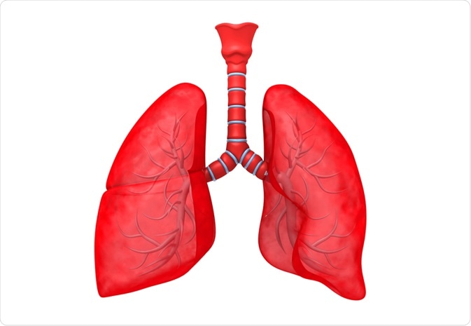
Pneumonia is an inflammatory condition occurring within the alveolar sacs of the lungs. It is caused by a variety of infectious microorganisms, including bacteria, viruses, fungi, and mycoplasma.
Explode | Shutterstock
The most predominant symptom of pneumonia is a deep cough, which can produce mucus that is yellow or green in color and may be tinged with blood. Other symptoms include difficulty breathing, chest pain, fever, chills, muscle aches, headaches, and sometimes delirium or mental confusion.
In most cases, pneumonia is highly treatable; however, people with pneumonia-like symptoms should seek medical care, as untreated pneumonia can develop into a serious medical condition. In order to diagnose pneumonia, a medical practitioner will perform a series of tests.
These tests examine the efficiency of oxygen delivery to the body and try to determine the infectious agent, which is necessary for determining which agent-specific therapy will be prescribed. In addition to these tests, imaging also plays a vital role in establishing a diagnosis.
How is pneumonia detected?
A visit to the doctor will always begin with a complete physical exam and medical history. During the exam, the doctor will perform an initial assessment of breathing quality using a stethoscope.
A patient with pneumonia may exhibit wheezing, bubbling, or crackling noises when breathing. These sounds are formed as air tries to pass through and over lung secretions.
How is pneumonia diagnosed?
If the breathing quality, as detected by the stethoscope, indicates an infection, a doctor will typically order one or more imaging tests to visualize the interior of the lung. These are done in order to identify the areas of infection and assess the overall severity of the disease.
Most patients will initially have to undergo a chest x-ray. However, in severe cases, or in patients where initial treatment methods are not producing positive results, a computerized tomography (CT) scan may be performed, which generates more detailed diagnostic imagery.
An alternative technique, used in particular for patients who are hospitalized and/or not responding to antibiotics, is bronchoscopy. This is a procedure used to visualize the airways. It involves the insertion of a tube containing a camera down the airway and into the lung where inflamed tissue may be observed.
Other tests involved in the diagnosis of pneumonia
Respiration, which is the exchange of gases, is the main function of the lungs. In this process oxygen is received from the air we breathe to oxygenate our blood, while carbon dioxide, which is a waste gas produced by metabolism, is removed.
Pneumonia negatively impacts this exchange process, as the buildup of mucous secretions interferes with the adequate oxygenation of blood. To assess if the lungs are operating at optimal levels with regards to their gas exchange, an arterial blood gas (ABG) test may be done.
An ABG is a blood test where blood is drawn from an artery as opposed to a vein. The most commonly used artery is the radial artery, however, other arteries may be used when necessary.
The ABG gives a highly accurate clinical representation of the patient’s respiratory status. In contrast to the ABG, pulse oximetry may also be done and although it is less invasive and painless, it is not as accurate as the ABG. Pulse oximetry involves the use of a small apparatus that may be attached to the finger to estimate blood oxygen levels.
How is the causative agent identified?
Given that pneumonia can be caused by one of over 30 different microorganisms, it is important to identify the underlying cause of each patient’s pneumonia, if possible. Distinguishing the causative agent is important for guiding subsequent treatment decisions. For example, bacterial-induced pneumonia can be treated with antibiotics, while fungal-induced pneumonia must be treated with antimycotics.
Testing can be done in a number of ways, including a sputum (mucous-based secretion brought up from a deep cough) test, a blood test, or a pleural fluid (liquid around the lining of the lungs and within the chest cavity) test. In each test, the substance or fluid is examined by microscope and/or cultured to identify causative organisms.
Sources
Further Reading
Last Updated: Nov 23, 2018


.png)


No hay comentarios:
Publicar un comentario