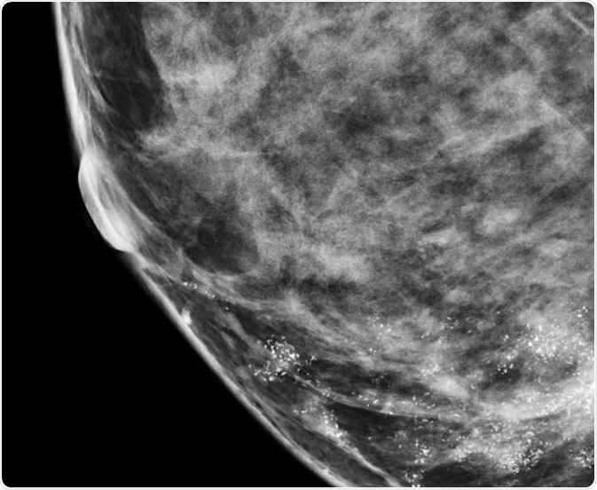
Microcalcifications in Breast Cancer
Calcifications in the breast can be benign or malignant. They can appear as either macrocalcifications or microcalcifications on a mammogram (i.e. an X-ray of the breast). Macrocalcifications look like large white dashes or dots and are mostly noncancerous and no further tests are required usually.

Microcalcifications in Breast
Microcalcifications are a reliable early sign of breast cancer and can be detected with the help of mammography. Microcalcifications can also give an idea of the extent of the disease. They are usually noncancerous, although some patterns can be a sign of cancer.
Information about the size, density, and distribution of breast microcalcifications can give an idea about the benign or malignant nature of the cancer. Also, more and more studies are linking mammary microcalcifications to lymph node invasion, HER-2 positivity, as well as poor prognosis for some patients with breast cancer.
The Importance of Microcalcifications
Microcalcifications are actually calcium deposits and are seen as tiny, white dots on a mammogram. They are much less common and are mostly a result of mutations in the breast tissue, though they can be caused by other factors. The appearance of microcalcifications is widely used in the detection of breast cancer at an early stage and can lead to better outcomes.
Nearly 50% of non-palpable cancers in the breast are detected only by the presence of microcalcifications on a mammogram, and approximately 93% of people with ductal carcinoma in situ (DCIS) also present with microcalcifications. Microcalcifications in the breast are very common in women and some studies have observed significant variations in the elemental composition of benign and malignant calcifications.
Types of Breast Microcalcifications
Breast microcalcifications can be divided into many types based on their form, size, density, and distribution.
Breast microcalcifications can occur in many different shapes or forms. They can be linear, round, granular, coarse, monomorphic when all of them having the same shape, or pleomorphic when several different shapes are present. They can also form clusters or groups.
When size is considered, breast microcalcifications can be either large or small, or in clusters with a mix of small and large microcalcifications. Furthermore, breast microcalcifications can have low, high, or variable density. When they are clustered, their homogeneous or non-homogeneous nature should be noted.
Finally, the distribution of breast microcalcifications refers to their placement on the mammogram. Microcalcifications can be unilateral or bilateral, multifocal, segmental, linear, diffuse, or regional.
Benign, Suspicious or Malignant Breast Microcalcifications
Several factors must be taken into account while deciding if a lesion is benign or malignant. Radiologists rely on breast microcalcifications as a possible indicator for breast cancer; however, only histological analysis can confirm this. When studying microcalcifications, if different findings lead to the same result (benign or malignant), then those predictions can be considered reliable.
In general, when the distribution of microcalcifications is diffuse or bilateral, when they have a round and/or punctuate shape, or when they are scattered in the breast tissue, the condition is usually benign. On the other hand, if microcalcifications have a linear or branching pattern with irregular borders, or if they have variable density, or when they are distributed in a haphazard or segmental way, there is a high probability that it is ductal carcinoma in situ or malignant cancer.
The nature of microcalcifications cannot always lead to a conclusive diagnosis and in such cases they are said to be ‘suspicious‘ – thus, a biopsy is required to confirm the benign or malignant nature of the cancer. If the distribution of the microcalcifications is linear and they are in round, oval, or amorphous form, they are termed as ‘suspicious’.
Radiologists usually term a variable distribution density where the breast microcalcifications are closely packed at one place and widely spread at another as ‘suspicious’, and not definitive, for cancer. It is referred to as ‘cluster of suspicious-looking microcalcifications0 on a mammogram report.
Benign lobular calcifications are usually round in shape and have a relatively higher density. They normally have pearl-like or well-defined contours and smooth borders. If the acinar lumen are small, they look punctate and have tiny spots on them.
Sources
Further Reading
Last Updated: Aug 23, 2018


No hay comentarios:
Publicar un comentario