Journal of Cardiovascular Magnetic Resonance
Automated cardiovascular magnetic resonance image analysis with fully convolutional networks
- Wenjia Bai,
- Matthew Sinclair,
- Giacomo Tarroni,
- Ozan Oktay,
- Martin Rajchl,
- Ghislain Vaillant,
- Aaron M. Lee,
- Nay Aung,
- Elena Lukaschuk,
- Mihir M. Sanghvi,
- Filip Zemrak,
- Kenneth Fung,
- Jose Miguel Paiva,
- Valentina Carapella,
- Young Jin Kim,
- Hideaki Suzuki,
- Bernhard Kainz,
- Paul M. Matthews,
- Steffen E. Petersen,
- Stefan K. Piechnik,
- Stefan Neubauer,
- Ben Glocker and
- Daniel Rueckert
Journal of Cardiovascular Magnetic Resonance201820:65
© The Author(s) 2018
- Received: 23 November 2017
- Accepted: 20 June 2018
- Published: 14 September 2018
Abstract
Background
Cardiovascular resonance (CMR) imaging is a standard imaging modality for assessing cardiovascular diseases (CVDs), the leading cause of death globally. CMR enables accurate quantification of the cardiac chamber volume, ejection fraction and myocardial mass, providing information for diagnosis and monitoring of CVDs. However, for years, clinicians have been relying on manual approaches for CMR image analysis, which is time consuming and prone to subjective errors. It is a major clinical challenge to automatically derive quantitative and clinically relevant information from CMR images.
Methods
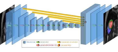
Deep neural networks have shown a great potential in image pattern recognition and segmentation for a variety of tasks. Here we demonstrate an automated analysis method for CMR images, which is based on a fully convolutional network (FCN). The network is trained and evaluated on a large-scale dataset from the UK Biobank, consisting of 4,875 subjects with 93,500 pixelwise annotated images. The performance of the method has been evaluated using a number of technical metrics, including the Dice metric, mean contour distance and Hausdorff distance, as well as clinically relevant measures, including left ventricle (LV) end-diastolic volume (LVEDV) and end-systolic volume (LVESV), LV mass (LVM); right ventricle (RV) end-diastolic volume (RVEDV) and end-systolic volume (RVESV).
Results
By combining FCN with a large-scale annotated dataset, the proposed automated method achieves a high performance in segmenting the LV and RV on short-axis CMR images and the left atrium (LA) and right atrium (RA) on long-axis CMR images. On a short-axis image test set of 600 subjects, it achieves an average Dice metric of 0.94 for the LV cavity, 0.88 for the LV myocardium and 0.90 for the RV cavity. The mean absolute difference between automated measurement and manual measurement is 6.1 mL for LVEDV, 5.3 mL for LVESV, 6.9 gram for LVM, 8.5 mL for RVEDV and 7.2 mL for RVESV. On long-axis image test sets, the average Dice metric is 0.93 for the LA cavity (2-chamber view), 0.95 for the LA cavity (4-chamber view) and 0.96 for the RA cavity (4-chamber view). The performance is comparable to human inter-observer variability.
Conclusions
We show that an automated method achieves a performance on par with human experts in analysing CMR images and deriving clinically relevant measures.
Keywords
- CMR image analysis
- Fully convolutional networks
- Machine learning


.png)


No hay comentarios:
Publicar un comentario