Neuroanatomy Model Systems
Neuroanatomy is the study of the relationship between structure and function in the brain and nervous system. One of the ways that neuroanatomy studies are carried out is by using other organisms as model systems. Caenorhabditis elegans (C. elegans), mice, zebrafish, and the fruit fly are common model systems used for neuroanatomy studies.
C. elegans - a model nematode
The nematode C. elegans has remarkable similarity to humans at the genetic level. About 40 percent of human disease genes have homologs in the C. elegans genome, and yet the organism is extremely simple, with only 1000 somatic cells. It regenerates quickly, and matures from egg to larva to fertile adult in just 3 days. C. elegans is also transparent, allowing it to be viewed on the inside in real time using light or fluorescent microscopy.
The nervous system of C. elegans is very simple, with only 302 neurons. The neurons join at the anterior of the worm in a nerve ring, a sort of primitive brain. The connections of those neurons have been mapped into a complete wiring diagram. C. elegans has simple behaviors like responses to touch and movement toward food that can be studied.
Fruit fly
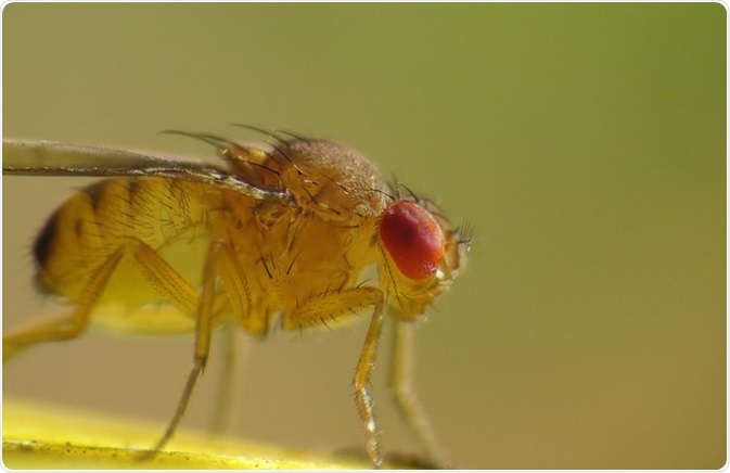
Drosophilia melanogaster, the fruit fly, is an organism whose biology and genetics have been extensively studied. Seventy-five percent of human disease-associated genes are represented in the Drosophila genome by functional homologs. It shares many organizational features of the mammalian central nervous system, yet has 1000 to 10,000 fewer neurons.

Image: Drosophila immigrans. Author: John Tann/ flickr.com. Shared under license.
Drosophila larvae have even fewer (no more than 3000 neurons) and they have become popular for use as a simple model system for the neuronal basis of higher brain functions and innate behaviors. Through previous studies of olfaction, gustation, and other sensory systems, the sensory networks of Drosophila are well-defined.
Mouse
Transgenic mouse modeling has become a staple for genetic neuroanatomy studies. The ability to change a single gene and observe the outcome is invaluable in genetic research, and being a mammal, mouse neuroanatomy has many valuable similarities to human neuroanatomy.
The main downside of using transgenic mice in neuroanatomy studies is that single gene alterations rarely model the full extent or presentation of the disease in humans. In addition, mice also have very small brains (compared to large human brains), which can be a drawback in some studies. Differences in neuroanatomy between human and mouse brain make it difficult to model some diseases of the central nervous system.
Zebrafish
The zebrafish (Danio rerio) is a common model system in biological science. It is a tropical freshwater fish of the minnow family native to the Himalayas. The structure of the central nervous system in fish has the same domains as the mammalian central nervous system, and the same neurotransmitters. Many genes connected to human neurodegenerative disease have been found in the zebrafish and are well characterized.

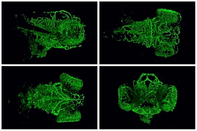
Image:5-day-old zebrafish heads showing development of blood vessels within the brain and eye. Author: NICHD/ /M. Butler/ flickr.com. Shared under license.
The zebrafish was among the first vertebrates to be cloned, and it has an ability to regenerate that makes it an attractive option as a model system. Another advantage of the zebrafish is its rapid development in the embryonic stage, and transparency of the embryos. They are also easy to handle and cost effective.
Sources
- https://www.ncbi.nlm.nih.gov/pmc/articles/PMC1855192/
- https://academic.oup.com/hmg/article-lookup/doi/10.1093/hmg/ddw428
- journals.plos.org/plosone/article?id=10.1371/journal.pone.0047518
- https://www.ncbi.nlm.nih.gov/pmc/articles/PMC3337025/
- www.sciencedirect.com/science/article/pii/S0969996110001610?via%3Dihub
Further Reading
Last Updated: Aug 23, 2018


No hay comentarios:
Publicar un comentario