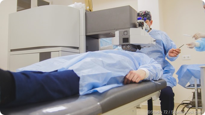
Small Incision Lenticule Extraction (SMILE) Surgery
Small incision lenticule extraction (SMILE) is a technique used in ocular surgery to treat an array of refractive errors. Errors of refraction that are corrected with the use of SMILE include hyperopia (i.e. farsightedness), myopia (i.e. shortsightedness), presbyopia (i.e. old-age long-sightedness) and astigmatism (i.e. imperfect corneal curvature). The SMILE technique was developed after another procedure known as the Femtosecond Lenticule Extraction (FLEX), which was an alternative to the laser-assisted in situ keratomileusis(LASIK).

In contrast to FLEX, SMILE allows for the extraction of the entire corneal lenticule without the creation of a flap being necessary. SMILE is a relatively new surgical operation. However, it is notable for some promising advantages in comparison to both LASIK and FLEX. While SMILE has an efficacy similar to the aforementioned two techniques, it has the potential for faster recovery times and re-innervation of the nerves within the cornea.
The SMILE procedure
Topical anesthetics are used prior to the start of the SMILE surgery. A femtosecond laser is used and the patient is raised to the level of the contact glass of the laser. Contact glasses are curved and calibrated individually and when the cornea makes contact with a contact glass, there is the appearance of a meniscus tear film that allows the patient to see fixation targets clearly. This is because fixation beam vergences are tailored to the refraction of patients.
Surgeons are able to activate suction ports at the point of contact on the cornea that fixate the eye. This allows for the auto-centering of the visual axis between the vertices of the cornea and the contact glass, and the lenticule that is being created. An Atlas topographical scan is used to confirm that centration of the axis has been acquired. If the visual axis is inadequately positioned, then the docking procedure is repeated after first releasing the suction.
The intrastromal lenticule’s lower interface is created prior to the subsequent creation of the upper interface or cap, which is then followed by a tunnel incision used to link the cap to the surface of the cornea. Once this is completed, a surgical microscope is used for separating the lenticule and for the extraction segment of the operation.
The surgeon opens the small incision and delineates the lower and upper edges of the lenticule in order to define tissue planes. There are numerous interface-separating instruments that may be used and the lenticule is removed with the help of a micro-retinal forceps once both layers have been separated.
Towards the end of the operation, uneven segments of the cap are evenly distributed to the periphery using a micro-spear, which is dry to avoid micro-folds. These may arise due to mismatching in length between the cap and bed after the extraction of the lenticule.
Complications
Although infrequently reported, complications, as with any other procedure, may arise. SMILE is a fairly safe and predictable operation. Problems such as abrasions of the epithelium, cap perforation and minute tears at the point of incision may occur.
References
- http://bjo.bmj.com/content/95/3/335?ijkey=ca9d3854b5f366b59548b570327ff2c1df05bb6c&keytype2=tf_ipsecsha
- http://eyewiki.aao.org/Small_Incision_Lenticule_Extraction_(SMILE)
- https://eandv.biomedcentral.com/articles/10.1186/s40662-014-0003-1
Further Reading
Last Updated: Aug 23, 2018


No hay comentarios:
Publicar un comentario