Types of Eye Allergy
Ocular allergies are both frequent and troublesome. The eye is often affected by allergic reactions of the eyelids, the conjunctiva and the cornea, either simultaneous with allergic rhinitis or by themselves. In fact, about 40 – 60% of allergic individuals have ocular symptoms.

Allergic conjunctivitis.
Group 1: Atopic conjunctivitis
These form the vast majority of ocular allergies, and are Immunoglobulin E- (IgE) mediated. They occur as a consequence of hypersensitivity to airborne pollutants, such as pollen grains, animal dander, mites and mold spores. Many of these people have allergic rhinitis.
Seasonal allergic conjunctivitis or hay fever conjunctivitis
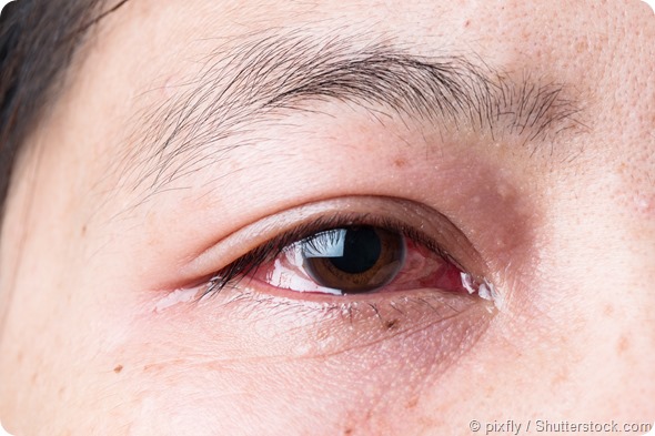
This is an acute or subacute illness due to pollen hypersensitivity. It is characterized by peaks and troughs, but becomes persistent during the allergy-inducing pollen/hay fever season. It manifests as itching, redness and swelling of the eyelids, with a burning sensation, chemosis (i.e. swelling of the conjunctiva), and excessive tear and mucus production.
It is treated with antihistamines, anti-inflammatory drugs and mast cell inhibitors, such as cromoglycate. Immunotherapy is sometimes used in severe cases. The aim of treatment is to completely eradicate the hypersensitivity reaction. This is attempted in order to prevent chronic inflammation, as well as treating other allergic manifestations, such as rhinitis.
Perennial allergic conjunctivitis
This condition mostly affects middle-aged people. It is linked to dry eye symptoms. Here the eye remains red and swollen and the patient feels a burning sensation of various degrees of severity, along with constant low-level pruritus (i.e. itching) of the eye.
This type occurs the whole year around and is thought to be due to dust mites, mold or dander. Treatment is with antihistamines, but only for a short period, because they worsen dryness. Steroids and mast cell inhibitors may also be used. These medications are usually required the whole year around.
Treatment should be prompt, intensive and prolonged if necessary. In both types, the affected individuals may have dark circles under their eyes. These marks are called allergy shiners. Such people may not tolerate bright light.
Group 2
These are more chronic, more serious and far less frequent, and may have corneal complications:
Vernal keratoconjunctivitis
Seen mostly in warm climates, this condition affects children or young adults. In about 50% of patients, other allergies are also present. It is seasonal in incidence, cropping up from the start of spring to fall.
In very hot climates, it may persist throughout the year. Affected people feel as if there is something in their eyes (i.e. a foreign body sensation), along with severe itching. Photophobia and the presence of thick mucus, with eosinophil granulomas in the upper eyelid, are other features.
It is thought to be a hypersensitivity reaction to airborne antigens. Both genetic and environmental factors are implicated in this condition. The incidence declines sharply over the age of 15 years. It is more frequent in males up to this age and in almost all patients it is bilateral. Three out of four patients also have a history of eczema or asthma.
Vernal keratoconjunctivitis is characterized by a very noticeable infiltration of inflammatory cells, which sets it apart from the conditions in group 1. Both T-helper (Th) cells type 1 and 2 are abundant along with their interleukin (IL) cytokines.
Th-1 cells are overexpressed especially in the stage of acute inflammation. In response to the massive release of chemical mediators, eosinophil infiltration and activation occurs, resulting in the elevation of eosinophil-derived epithelial toxins. This causes corneal damage in many cases. It is usually in the form of punctate keratitis, leading to the formation of corneal plaques and ulcers.
In addition, Th-2 cytokines IL-4 and IL-13 stimulate the production of collagen by inducing fibroblast migration, proliferation and collagen synthesis. The typical giant papillae are formed by the over-activity of matrix metalloproteinases released by fibroblasts, which induce extracellular matrix deposition.
An overall imbalance between stimulatory and inhibitory activity leads to prolonged inflammation. Treatment is with antihistamines and mast cell inhibitors. Cyclosporine A and corticosteroids are used in severe disease.
Atopic keratoconjunctivitis
Unlike vernal keratoconjunctivitis, this is part of an abnormally sensitive immune reaction, often accompanied by atopic dermatitis, allergic rhinitis or asthma. It occurs over a wide level of severity and may threaten eyesight.
Symptoms include burning, severe itching and redness of the eyes, with thick mucus, which may gum together the eyelids after sleep. Eczematous blepharitis is present, often with Staphylococcus aureus secondary infection.
The eyelashes often fall out. Corneal damage is likely if untreated. Blepharitis is treated with the calcineurin inhibitors tacrolimus or pimecrolimus until it resolves completely. Corticosteroids and, in some cases, cyclosporine A, is required to treat the conjunctivitis. Cromoglycate and antihistamines are also administered.
Group 3
These are often not considered to be real allergies, but are caused by repeated micro-traumas of the cornea.
Contact allergic hypersensitivity
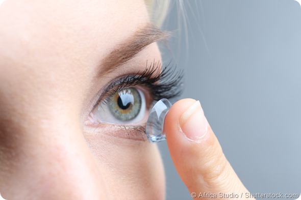
Contact lenses may induce allergy by themselves or because of antigenic tear protein-lens combinations. Symptoms include redness, itching and mucus production, with significant discomfort on wearing the lens.

Some eye allergies can be caused by contact lenses.
Giant papillary conjunctivitis
This form is also due to contact lenses. Large papules or vesicles form on the inner conjunctiva of the upper eyelid. The eyes are puffy, itchy and teary. There is discomfort on wearing contact lenses and mucus production. A foreign body sensation is present, while vision may be blurred.
Group 4
These are caused by immune and toxic damage to the surface of the eyes, due to contact with various substances.
Contact blepharoconjunctivitis
This is a T-cell mediated hypersensitivity reaction, which is caused by the binding of haptens (i.e. small molecules that can elicit antibody production) to tissue proteins. The haptens may be natural substances, such as natural resin in cosmetics, or nickel and cobalt in mascara, and components of eye medications.
The offending substance may be an antibiotic, glaucoma medication, antihistamines or mydriatics (i.e. agents that induce pupil dilation). Sometimes even the preservatives used in these topical medications may cause allergy or inflammation. Treatment is by preservative-free artificial tears and antihistamines as required.
Delayed allergy
This may be caused by airborne pollutants, chemicals, such as those vaporized from rubber or composites, or some plants in the hot humid conditions of a greenhouse. Treatment is by avoiding the offending chemical and by treating with corticosteroids, tacrolimus or pimecrolimus.
References
- https://www.ncbi.nlm.nih.gov/pmc/articles/PMC2515354/
- http://www.ncbi.nlm.nih.gov/pmc/articles/PMC3047907/
- http://acaai.org/allergies/types/eye-allergies
Further Reading
Last Updated: Aug 23, 2018


No hay comentarios:
Publicar un comentario