Los avances de la medicina en el campo de la genética, por ende de la herencia, están modificando el paisaje del conocimiento médico de las enfermedades. Este BLOG intenta informar acerca de los avances proveyendo orientación al enfermo y su familia así como información científica al profesional del equipo de salud de habla hispana.
martes, 5 de marzo de 2019
Breast Cancer Treatment (PDQ®) 1/3 —Patient Version - National Cancer Institute
Breast cancer is a disease in which malignant (cancer) cells form in the tissues of the breast.
A family history of breast cancer and other factors increase the risk of breast cancer.
Breast cancer is sometimes caused by inherited gene mutations (changes).
The use of certain medicines and other factors decrease the risk of breast cancer.
Signs of breast cancer include a lump or change in the breast.
Tests that examine the breasts are used to detect (find) and diagnose breast cancer.
If cancer is found, tests are done to study the cancer cells.
Certain factors affect prognosis (chance of recovery) and treatment options.
Breast cancer is a disease in which malignant (cancer) cells form in the tissues of the breast.
The breast is made up of lobes and ducts. Each breast has 15 to 20 sections called lobes. Each lobe has many smaller sections called lobules. Lobules end in dozens of tiny bulbs that can make milk. The lobes, lobules, and bulbs are linked by thin tubes called ducts.
ENLARGEAnatomy of the female breast. The nipple and areola are shown on the outside of the breast. The lymph nodes, lobes, lobules, ducts, and other parts of the inside of the breast are also shown.
Each breast also has blood vessels and lymph vessels. The lymph vessels carry an almost colorless, watery fluid called lymph. Lymph vessels carry lymph between lymph nodes. Lymph nodes are small, bean-shaped structures found throughout the body. They filterlymph and store white blood cells that help fight infection and disease. Groups of lymph nodes are found near the breast in the axilla (under the arm), above the collarbone, and in the chest.
The most common type of breast cancer is ductal carcinoma, which begins in the cells of the ducts. Cancer that begins in the lobes or lobules is called lobular carcinoma and is more often found in both breasts than are other types of breast cancer. Inflammatory breast cancer is an uncommon type of breast cancer in which the breast is warm, red, and swollen.
See the following PDQ summaries for more information about breast cancer:
A family history of breast cancer and other factors increase the risk of breast cancer.
Anything that increases your chance of getting a disease is called a risk factor. Having a risk factor does not mean that you will get cancer; not having risk factors doesn't mean that you will not get cancer. Talk to your doctor if you think you may be at risk for breast cancer.
Risk factors for breast cancer include the following:
Older age is the main risk factor for most cancers. The chance of getting cancer increases as you get older.
NCI'sBreast Cancer Risk Assessment Tool uses a woman's risk factors to estimate her risk for breast cancer during the next five years and up to age 90. This online tool is meant to be used by a health care provider. For more information on breast cancer risk, call 1-800-4-CANCER.
Breast cancer is sometimes caused by inherited gene mutations (changes).
The genes in cells carry the hereditary information that is received from a person’s parents. Hereditary breast cancer makes up about 5% to 10% of all breast cancer. Some mutated genes related to breast cancer are more common in certain ethnic groups.
Women who have certain gene mutations, such as a BRCA1 or BRCA2 mutation, have an increased risk of breast cancer. These women also have an increased risk of ovarian cancer, and may have an increased risk of other cancers. Men who have a mutated gene related to breast cancer also have an increased risk of breast cancer. For more information, see the PDQ summary on Male Breast Cancer Treatment.
There are tests that can detect (find) mutated genes. These genetic tests are sometimes done for members of families with a high risk of cancer. See the PDQ summary on Genetics of Breast and Gynecologic Cancers for more information.
The use of certain medicines and other factors decrease the risk of breast cancer.
Anything that decreases your chance of getting a disease is called a protective factor.
Protective factors for breast cancer include the following:
Fluid, other than breast milk, from the nipple, especially if it's bloody.
Scaly, red, or swollen skin on the breast, nipple, or areola (the dark area of skin around the nipple).
Dimples in the breast that look like the skin of an orange, called peau d’orange.
Tests that examine the breasts are used to detect (find) and diagnose breast cancer.
Check with your doctor if you notice any changes in your breasts. The following tests and procedures may be used:
Physical exam and history: An exam of the body to check general signs of health, including checking for signs of disease, such as lumps or anything else that seems unusual. A history of the patient’s health habits and past illnesses and treatments will also be taken.
Clinical breast exam (CBE): An exam of the breast by a doctor or other health professional. The doctor will carefully feel the breasts and under the arms for lumps or anything else that seems unusual.
Mammogram: An x-ray of the breast.ENLARGEMammography. The breast is pressed between two plates. X-rays are used to take pictures of breast tissue.
Ultrasound exam: A procedure in which high-energy sound waves (ultrasound) are bounced off internal tissues or organs and make echoes. The echoes form a picture of body tissues called a sonogram. The picture can be printed to be looked at later.
MRI (magnetic resonance imaging): A procedure that uses a magnet, radio waves, and a computer to make a series of detailed pictures of both breasts. This procedure is also called nuclear magnetic resonance imaging (NMRI).
Blood chemistry studies: A procedure in which a blood sample is checked to measure the amounts of certain substances released into the blood by organs and tissues in the body. An unusual (higher or lower than normal) amount of a substance can be a sign of disease.
Biopsy: The removal of cells or tissues so they can be viewed under a microscope by a pathologist to check for signs of cancer. If a lump in the breast is found, a biopsy may be done.
There are four types of biopsy used to check for breast cancer:
Multigene tests: Tests in which samples of tissue are studied to look at the activity of many genes at the same time. These tests may help predict whether cancer will spread to other parts of the body or recur (come back).
There are many types of multigene tests. The following multigene tests have been studied in clinical trials:
Oncotype DX: This test helps predict whether early-stage breast cancer that is estrogen receptor positive and node negative will spread to other parts of the body. If the risk that the cancer will spread is high, chemotherapy may be given to lower the risk.
MammaPrint: This test helps predict whether early-stage breast cancer will spread to other parts of the body. If the risk that the cancer will spread is high, chemotherapy may be given to lower the risk.
Based on these tests, breast cancer is described as one of the following types:
Triple negative (estrogen receptor, progesterone receptor, and HER2/neu negative).
This information helps the doctor decide which treatments will work best for your cancer.
Certain factors affect prognosis (chance of recovery) and treatment options.
The prognosis (chance of recovery) and treatment options depend on the following:
The stage of the cancer (the size of the tumor and whether it is in the breast only or has spread to lymph nodes or other places in the body).
The type of breast cancer.
Estrogen receptor and progesterone receptor levels in the tumor tissue.
Human epidermal growth factor type 2 receptor (HER2/neu) levels in the tumor tissue.
Whether the tumor tissue is triple negative (cells that do not have estrogen receptors, progesterone receptors, or high levels of HER2/neu).
How fast the tumor is growing.
How likely the tumor is to recur (come back).
A woman’s age, general health, and menopausal status (whether a woman is still having menstrual periods).
Whether the cancer has just been diagnosed or has recurred (come back).
Stages of Breast Cancer
KEY POINTS
After breast cancer has been diagnosed, tests are done to find out if cancer cells have spread within the breast or to other parts of the body.
There are three ways that cancer spreads in the body.
Cancer may spread from where it began to other parts of the body.
In breast cancer, stage is based on the size and location of the primary tumor, the spread of cancer to nearby lymph nodes or other parts of the body, tumor grade, and whether certain biomarkers are present.
The TNM system is used to describe the size of the primary tumor and the spread of cancer to nearby lymph nodes or other parts of the body.
Tumor (T). The size and location of the tumor.
Lymph Node (N). The size and location of lymph nodes where cancer has spread.
Metastasis (M). The spread of cancer to other parts of the body.
The grading system is used to describe how quickly a breast tumor is likely to grow and spread.
Biomarker testing is used to find out whether breast cancer cells have certain receptors.
The TNM system, the grading system, and biomarker status are combined to find out the breast cancer stage.
Talk to your doctor to find out what your breast cancer stage is and how it is used to plan the best treatment for you.
The treatment of breast cancer depends partly on the stage of the disease.
After breast cancer has been diagnosed, tests are done to find out if cancer cells have spread within the breast or to other parts of the body.
The process used to find out whether the cancer has spread within the breast or to other parts of the body is called staging. The information gathered from the staging process determines the stage of the disease. It is important to know the stage in order to plan treatment. The results of some of the tests used to diagnosebreast cancer are also used to stage the disease. (See the General Information section.)
The following tests and procedures also may be used in the staging process:
Sentinel lymph node biopsy: The removal of the sentinel lymph node during surgery. The sentinel lymph node is the first lymph node in a group of lymph nodes to receive lymphatic drainage from the primary tumor. It is the first lymph node the cancer is likely to spread to from the primary tumor. A radioactive substance and/or blue dye is injected near the tumor. The substance or dye flows through the lymph ducts to the lymph nodes. The first lymph node to receive the substance or dye is removed. A pathologist views the tissue under a microscope to look for cancer cells. If cancer cells are not found, it may not be necessary to remove more lymph nodes. Sometimes, a sentinel lymph node is found in more than one group of nodes.
Chest x-ray: An x-ray of the organs and bones inside the chest. An x-ray is a type of energy beam that can go through the body and onto film, making a picture of areas inside the body.
CT scan (CAT scan): A procedure that makes a series of detailed pictures of areas inside the body, taken from different angles. The pictures are made by a computer linked to an x-ray machine. A dye may be injected into a vein or swallowed to help the organs or tissues show up more clearly. This procedure is also called computed tomography, computerized tomography, or computerized axial tomography.
Bone scan: A procedure to check if there are rapidly dividing cells, such as cancer cells, in the bone. A very small amount of radioactive material is injected into a vein and travels through the bloodstream. The radioactive material collects in the bones with cancer and is detected by a scanner.
PET scan (positron emission tomography scan): A procedure to find malignanttumor cells in the body. A small amount of radioactive glucose (sugar) is injected into a vein. The PET scanner rotates around the body and makes a picture of where glucose is being used in the body. Malignant tumor cells show up brighter in the picture because they are more active and take up more glucose than normal cells do.
There are three ways that cancer spreads in the body.
Tissue. The cancer spreads from where it began by growing into nearby areas.
Lymph system. The cancer spreads from where it began by getting into the lymph system. The cancer travels through the lymph vessels to other parts of the body.
Blood. The cancer spreads from where it began by getting into the blood. The cancer travels through the blood vessels to other parts of the body.
Cancer may spread from where it began to other parts of the body.
When cancer spreads to another part of the body, it is called metastasis. Cancer cells break away from where they began (the primary tumor) and travel through the lymph system or blood.
Lymph system. The cancer gets into the lymph system, travels through the lymph vessels, and forms a tumor (metastatic tumor) in another part of the body.
Blood. The cancer gets into the blood, travels through the blood vessels, and forms a tumor (metastatic tumor) in another part of the body.
The metastatic tumor is the same type of cancer as the primary tumor. For example, if breast cancer spreads to the bone, the cancer cells in the bone are actually breast cancer cells. The disease is metastatic breast cancer, not bone cancer.
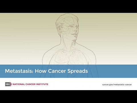
metastasis: how cancer spreads
Many cancer deaths are caused when cancer moves from the original tumor and spreads to other tissues and organs. This is called metastatic cancer. This animation shows how cancer cells travel from the place in the body where they first formed to other parts of the body.
ver historia personal en: www.cerasale.com.ar [dado de baja por la Cancillería Argentina por temas políticos, propio de la censura que rige en nuestro medio]//
www.revistamedicos.com.ar //
www.quorumtuc.com.ar //
www.sectorsalud.com.ar //
www.maimonides.edu //
weblog.maimonides.edu/farmacia/archives/UM_Informe_Autoevaluacion_FyB.pdf - //
weblog.maimonides.edu/farmacia/archives/0216_Admin_FarmEcon.pdf - //
www.documentalistas.org.ar //
www.cpcesfe2.org.ar //
www.nogracias.eu //
www.estenssorome.com.ar //
www.cuautitlan.unam.mx/descargas/licenciaturas/bqd/plandestudio_bqd_ //
www.latamjpharm.org/trabajos/25/2/LAJOP_25_2_6_1_M4M6Z9746D.pdf //
www.nogracias.eu/v_juventud/informacion/informacionver.asp?cod= //
www.colfarse.com.ar //
www.proz.com/kudoz/english_to_spanish/art_literary/523942-key_factors.html - 65k - // www.llave.connmed.com.ar/portalnoticias_vernoticia.php?codigonoticia=17715 // www.frusculleda.com.ar/homepage/espanol/activities_teaching.htm // http://www.on24.com.ar/nota.aspx?idNot=36331 ||


.png)


No hay comentarios:
Publicar un comentario