Kidney Stones - Treatment
The treatment decision for kidney stones is based on the stone type, severity of the condition, and duration of the symptoms in patients. Though plenty of treatment options are available, the procedure that best suits an individual should be discussed to help improving efficacy of treatment and patient outcomes.
Non-Surgical Procedure
A waiting period of about 4–6 weeks is considered to be safe for the stones to pass on their own, provided the patients can bear the pain and symptoms of infection are not visible. When the size of the stone is small, it does not block the kidney completely and it may pass in the urine naturally.
Drinking sufficient water and using medicines for pain relief and nausea are found to be helpful. The ureters can be relaxed by using medications that help the stone to move out easily.
Surgical Procedure
When the stone is large, it may not pass through easily or the pain can be unbearable until the stone is passed out. When the stone causes repetitive urinary infection or when it blocks the urine flow, a surgical procedure may be recommended.
In general, there are three types of surgeries available for removing kidney stones. They are:
- Shock wave lithotripsy
- Ureteroscopy
- Percutaneous lithotripsy
Anesthesia is given to patients before performing the surgery to make them comfortable during the surgery.

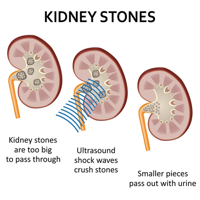
Shock waves used to break a kidney stone into small pieces that can more easily travel through the urinary tract and pass from the body. Imaged Credit: Neokryuger / Shutterstock
Shock Wave Lithotripsy
In shock wave lithotripsy (SWL) treatment, ultrasound or X-ray are used to direct shock waves to target the stone accurately. The stone is broken down into tiny pieces when shock waves are repeatedly fired at the stone so that smaller stone pieces eventually come out when the patient passes urine.
The stones made up of calcium phosphate or calcium oxalate are hard, similar to cystine stones. In such cases, this type of surgery is not suitable. Moreover, when the stone is very large, SWL is not suggested.
Though considered to be a very safe procedure, SWL has side effects as well. For a few days after surgery, patients can notice blood in their urine. Although most of the stone pieces are discharged painlessly, bigger pieces get caught in the ureter, which can cause pain and physicians may recommend other stone removal methods for this reason.
Patients are usually discharged on the same day of the surgery and can resume normal activities within 2–3 days. A strainer is used for collecting pieces of stones that are passed out in the urine and sent to a laboratory to find out the cause of stone formation.
Ureteroscopy
In ureteroscopy (URS), a ureteroscope—a small telescope—is inserted into the kidney through the bladder. When stones are found in the lower portion of the ureter near the bladder, rigid telescopes are employed. On the other hand, when stones are seen in the upper part of the ureter and kidney, flexible telescopes are preferred.
Using this equipment, urologists can view the stones without cutting them. A device resembling a small basket is used to collect small stones from the ureter or kidney. In the case of larger stones, other tools that are capable of breaking them, such as laser, are used; the stones are broken down into smaller pieces.
After the removal of stones, a small, rigid, plastic tube, called a stent, is placed temporarily in the ureter. The stent keeps the ureter open and assists in draining urine from the kidney to the bladder. As the stent resides in the body, no separate external bag is required for urine collection.
Physicians remove the stent 4–10 days after the surgery. There are also provisions for the patients to remove the stent by themselves. However, it is advisable to remove the stent according to the direction of the physician. Using the stent for a longer duration could cause an infection and can even disturb the function of the kidney.
Percutaneous Lithotripsy
When the stones are dense or too many in number or bigger in size, say above 2 cm, percutaneous lithotripsy (PCNL) is performed. Compared with SWL or URS, PCNL is a more invasive procedure. In the side or back of the kidney, a cut of about half inch is made. A nephroscope—a rigid telescope—is inserted into the incision to reach the stone. The stone is broken down into smaller pieces and sucked by an instrument in the nephroscope. After completing the surgery, the physician inserts a tube in the kidney, which helps to drain the urine into a pouch placed externally to prevent bleeding.
Patients who undergo percutaneous lithotripsy require an overnight stay in the hospital. Urologists may perform X-rays to check if any stone pieces are left over. If stones are still present, the surgery is performed again. The tube can be removed after a couple of days and individuals can resume their usual routine after 1–2 weeks.
Rare surgeries such as robotic surgery or laparoscopic surgery are performed for kidney stone removal when other treatment methods do not yield results.
Sources
Further Reading
Last Updated: Feb 26, 2019


No hay comentarios:
Publicar un comentario