hereditary leiomyomatosis and renal cell cancer
Hereditary leiomyomatosis and renal cell cancer (HLRCC) is a disorder in which affected individuals tend to develop benign tumors containing smooth muscle tissue (leiomyomas) in the skin and, in females, the uterus. This condition also increases the risk of kidney cancer.
In this disorder, growths on the skin (cutaneous leiomyomas) typically develop in the third decade of life. Most of these growths arise from the tiny muscles around the hair follicles that cause "goosebumps". They appear as bumps or nodules on the trunk, arms, legs, and occasionally on the face. Cutaneous leiomyomas may be the same color as the surrounding skin, or they may be darker. Some affected individuals have no cutaneous leiomyomas or only a few, but the growths tend to increase in size and number over time. Cutaneous leiomyomas are often more sensitive than the surrounding skin to cold or light touch, and may be painful.
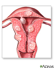
Most women with HLRCC also develop uterine leiomyomas (fibroids). While uterine fibroids are very common in the general population, women with HLRCC tend to have numerous large fibroids that appear earlier than in the general population.
Approximately 10 percent to 16 percent of people with HLRCC develop a type of kidney cancer called renal cell cancer. The signs and symptoms of renal cell cancer may include lower back pain, blood in the urine, or a mass in the kidney that can be felt upon physical examination. Some people with renal cell cancer have no symptoms until the disease is advanced. The average age at which people with HLRCC are diagnosed with kidney cancer is in their forties.
This disorder, especially if it appears in individuals or families without renal cell cancer, is also sometimes called multiple cutaneous leiomyomatosis (MCL) or multiple cutaneous and uterine leiomyomatosis (MCUL).

Uterine Fibroids Update
MedlinePlus sent this bulletin at 04/18/2016 01:11 PM EDTNew on the MedlinePlus Uterine Fibroids page:
04/13/2016 02:39 PM EDT
Source: National Library of Medicine - 
National Institutes of Health
NIH MedlinePlus Magazine
Uterine fibroids are the most common benign tumors in women of childbearing age. Fibroids are made of muscle cells and other tissues that grow in and around the wall of the uterus, or womb. The cause of fibroids is unknown. Risk factors include being African American or being overweight.
Many women with fibroids have no symptoms. If you do have symptoms, they may include
- Heavy or painful periods or bleeding between periods
- Feeling "full" in the lower abdomen
- Urinating often
- Pain during sex
- Lower back pain
- Reproductive problems, such as infertility, multiple miscarriages or early labor
Your health care provider may find fibroids during a gynecological exam or by using imaging tests. Treatment includes drugs that can slow or stop their growth, or surgery. If you have no symptoms, you may not even need treatment. Many women with fibroids can get pregnant naturally. For those who cannot, infertility treatments may help.
NIH: National Institute of Child Health and Human Development
- Uterine Fibroids (American College of Obstetricians and Gynecologists)
- Uterine Fibroids (National Institute of Child Health and Human Development)
- Uterine Fibroids Fact Sheet (Department of Health and Human Services, Office on Women's Health)Available in Spanish
- FDA OKs 'Containment' Bag for Certain Uterine Surgeries (04/07/2016, HealthDay)
- Women's Sex Lives Get a Boost After Non-Surgical Fibroid Treatment (04/04/2016, HealthDay)
- What Are the Symptoms of Uterine Fibroids? (National Institute of Child Health and Human Development)
- How Are Uterine Fibroids Diagnosed? (National Institute of Child Health and Human Development)
- Hysteroscopy (American College of Obstetricians and Gynecologists) - PDFAvailable in Spanish
- Pelvic laparoscopy - slideshow Available in Spanish
- Ultrasound -- Pelvis
 (Radiological Society of North America, American College of Radiology)Available in Spanish
- Focused Ultrasound Surgery for Uterine Fibroids (Mayo Foundation for Medical Education and Research)
- Medical Treatments for Fibroids (National Institute of Child Health and Human Development)
- Myomectomy (Mayo Foundation for Medical Education and Research)
- Surgical Treatments for Fibroids (National Institute of Child Health and Human Development)
- Uterine Artery Embolization (Mayo Foundation for Medical Education and Research)
- Uterine Fibroid Embolization (UFE) (Radiological Society of North America)Available in Spanish
- Genetics Home Reference: hereditary leiomyomatosis and renal cell cancer (National Library of Medicine)
- Uterine Fibroid Embolization (OR-Live) - South Miami Hospital, Miami, FL 3/25/2010Available in Spanish
- Genetic Cause of Infertility Associated with Uterine Fibroids (National Institute of Child Health and Human Development)
- ClinicalTrials.gov: Leiomyoma (National Institutes of Health)
- ClinicalTrials.gov: Uterine Myomectomy (National Institutes of Health)
- Find an Interventional Radiologist (Society of Interventional Radiology)
- National Institute of Child Health and Human Development Available in Spanish
- womenshealth.gov (Department of Health and Human Services, Office on Women's Health)Available in Spanish
- Hysteroscopy Available in Spanish
- Living with uterine fibroids Available in Spanish
- Uterine artery embolization Available in Spanish
- Uterine artery embolization - discharge Available in Spanish
- Uterine fibroids Available in Spanish


.png)


No hay comentarios:
Publicar un comentario